
2014-12-03
The Contribution of Optical Coherence Tomography to Interventional Cardiology
Publication
Publication
Methodological Considerations and Clinical Application
De bijdrage van optische coherentie tomografie in interventionele cardiologie: methodologische overwegingen en klinische toepassingen
Abstract
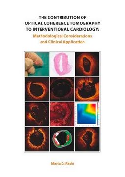
Since the beginning of interventional cardiology, coronary angiography has been the reference tool for assessing the severity of coronary lesions and guide stent implantation. With growing knowledge about the pathophysiology of atherothrombosis and stent failure it became of interest to visualise in vivo different processes taking place at the level of the coronary vessel wall for the purpose of improving cardiovascular outcomes. Intracoronary imaging techniques overcome the lumenographic limitations of angiography by enabling a histology-like cross-sectional view of the vessel wall and implanted devices. Although intravascular ultrasound (IVUS) has provided valuable insights into the dynamic nature of atherosclerosis and the causes of stent failure, the technology has a limited axial resolution (100-250 μm) and poor ability to differentiate between various tissue components. Intracoronary optical coherence tomography (OCT) has opened the door to a new world in interventional cardiology. With a nearhistological resolution of 10-20 μm, this near-infrared light-based technology offers a significantly improved visualisation of plaque and stent-related features. However, only after its introduction did we realise how much the technology differs from IVUS beyond the level of resolution: in terms of aspects related to the use of light rather than sound, affecting tissue and stent characterisation and resulting in a unique array of artefacts; but also as a consequence of the significantly increased amount of data and its hierarchical nature requiring an adjustment of the qualitative-, quantitative- and statistical methods for an appropriate utilisation of the technology. This thesis will provide an insight into the development of these methods and their clinical application.
| Additional Metadata | |
|---|---|
| , | |
| P.W.J.C. Serruys (Patrick) | |
| Erasmus University Rotterdam | |
| hdl.handle.net/1765/77217 | |
| Organisation | Erasmus MC: University Medical Center Rotterdam |
|
Radu, M. (2014, December 3). The Contribution of Optical Coherence Tomography to Interventional Cardiology. Retrieved from http://hdl.handle.net/1765/77217 |
|